Contents
- 💡 What is Computed Tomography (CT)?
- 🔬 How CT Scans Work: The Engineering Behind the Image
- 🏥 Who Needs a CT Scan? Applications and Indications
- 🆚 CT vs. Other Imaging Modalities: Making the Choice
- ⚡ The Vibe of CT: Cultural Resonance and Impact
- 💰 Cost and Accessibility: Navigating the Healthcare System
- ⚠️ Risks and Considerations: A Skeptic's View
- 🚀 The Future of CT: Innovations and Next Steps
- Frequently Asked Questions
- Related Topics
Overview
Computed Tomography (CT) revolutionized medical imaging since its inception in the early 1970s, with the first commercial scanner developed by Godfrey Hounsfield and Allan Cormack. This technology combines X-ray images taken from different angles to create cross-sectional views of the body, allowing for unprecedented diagnostic capabilities. The precision of CT scans has made them indispensable in emergency medicine, oncology, and surgical planning. However, the rise of CT also brings concerns over radiation exposure and cost. As artificial intelligence and machine learning integrate into imaging, the future of CT promises enhanced accuracy and efficiency, but raises ethical questions about data privacy and algorithmic bias.
💡 What is Computed Tomography (CT)?
Computed Tomography (CT), a sophisticated evolution of earlier computed axial tomography, stands as a cornerstone of modern medical diagnostics. It's a non-invasive imaging technique that generates detailed, cross-sectional views of the body's internal structures, far surpassing the capabilities of standard X-rays. Performed by highly trained radiographers or radiology technologists, a CT scan provides physicians with critical anatomical information for diagnosing a vast array of conditions, from bone fractures to complex tumors. Its ability to visualize soft tissues, blood vessels, and organs with remarkable clarity makes it indispensable in emergency medicine and routine diagnostic workups alike.
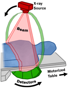
🔬 How CT Scans Work: The Engineering Behind the Image
At its heart, a CT scanner is a marvel of engineering. It employs an X-ray tube that rotates around the patient, emitting a fan-shaped beam of X-rays. Opposite the tube, a detector array measures how much the X-rays are attenuated (absorbed or scattered) by the various tissues they pass through. This process captures hundreds, even thousands, of individual measurements from multiple angles. These raw data points are then fed into powerful computers, where complex tomographic reconstruction algorithms—mathematical processes developed over decades—transform them into precise, slice-like images of the body. This intricate interplay of hardware and software allows for the creation of detailed anatomical maps.
🏥 Who Needs a CT Scan? Applications and Indications
The utility of CT scans spans a broad spectrum of medical needs. They are crucial for detecting and staging cancers, identifying internal injuries after trauma, diagnosing vascular diseases like aneurysms, and evaluating conditions affecting the brain, lungs, and abdomen. A significant advantage of CT is its compatibility with patients who have metallic implants or pacemakers, conditions that often preclude the use of Magnetic Resonance Imaging (MRI). This broad applicability ensures that CT remains a go-to modality for a wide range of clinical scenarios, from acute emergencies to chronic disease management.
🆚 CT vs. Other Imaging Modalities: Making the Choice
When selecting an imaging modality, understanding the distinctions between CT, X-ray, Ultrasound, and MRI is paramount. Standard X-rays offer quick, two-dimensional views, ideal for bone imaging but limited for soft tissues. Ultrasound uses sound waves, making it safe for pregnant patients and excellent for real-time imaging of organs and blood flow, though operator-dependent. MRI, while offering superior soft tissue contrast without ionizing radiation, can be time-consuming and is contraindicated for patients with certain metallic implants. CT strikes a balance, providing rapid, detailed cross-sectional imaging with good soft tissue and excellent bone visualization, making it a versatile choice for many diagnostic puzzles.
⚡ The Vibe of CT: Cultural Resonance and Impact
The cultural resonance of CT scans is profound, albeit often unseen by the public. It represents a significant leap in humanity's ability to peer inside the living body without surgery, a concept that would have seemed like science fiction just a century ago. The development of CT, pioneered by figures like Sir Godfrey Hounsfield, earned him a Nobel Prize in Physiology or Medicine in 1979, underscoring its monumental impact on medicine. While the technology itself is clinical, its existence has fostered a societal expectation of detailed internal visualization, influencing everything from medical dramas to patient understanding of their own health.
⚠️ Risks and Considerations: A Skeptic's View
Despite its immense benefits, it's crucial to acknowledge the inherent risks associated with CT scans, primarily the use of ionizing radiation. While the radiation dose from a single CT scan is generally considered safe and the benefits of accurate diagnosis often outweigh the risks, cumulative exposure over a lifetime is a concern. Patients with certain medical conditions or those undergoing frequent scans may warrant closer monitoring. Furthermore, the use of contrast agents—often administered intravenously to enhance visualization of blood vessels and tissues—can, in rare cases, lead to allergic reactions or kidney problems, necessitating careful patient screening.
🚀 The Future of CT: Innovations and Next Steps
The future of CT imaging is bright, driven by relentless innovation. We're seeing advancements in low-dose CT technology, significantly reducing radiation exposure without compromising image quality. Dual-energy CT allows for material decomposition, providing more information about tissue composition and aiding in the diagnosis of conditions like gout or kidney stones. Artificial intelligence (AI) is also playing an increasingly vital role, assisting in image reconstruction, automating lesion detection, and improving workflow efficiency. These developments promise to make CT scans even safer, more informative, and more accessible in the years to come.
Key Facts
- Year
- 1972
- Origin
- Developed by Godfrey Hounsfield and Allan Cormack
- Category
- Medical Imaging
- Type
- Technology
Frequently Asked Questions
Is a CT scan painful?
No, a CT scan is a painless procedure. You will lie on a table that slides into the CT scanner. The scanner itself is a large, donut-shaped machine that rotates around you to capture images. You may be asked to hold your breath for short periods during the scan to prevent motion blur. The only potential discomfort might come from the insertion of an IV line if contrast dye is being used.
How long does a CT scan take?
The actual scanning time for a CT is typically very fast, often lasting only a few minutes. However, the entire process, including preparation (like changing into a gown and IV placement if needed) and positioning, can take anywhere from 10 to 30 minutes. Complex scans or those requiring multiple sequences might extend this duration.
Can I eat or drink before a CT scan?
Generally, you can eat and drink normally before most CT scans. However, your doctor or the imaging facility will provide specific instructions. If a contrast agent is to be used, you might be asked to fast for a few hours beforehand to reduce the risk of nausea or vomiting. Always confirm dietary restrictions with your healthcare provider.
What is contrast dye in a CT scan?
Contrast dye, often iodine-based, is a special liquid that helps to highlight certain tissues, blood vessels, or abnormalities on the CT images. It can be administered orally (swallowed), rectally (as an enema), or intravenously (injected into a vein). The dye makes these areas appear brighter or more distinct on the scan, aiding in diagnosis.
Are CT scans safe for pregnant women?
CT scans expose patients to ionizing radiation, which can pose risks to a developing fetus. Therefore, CT scans are generally avoided in pregnant women unless absolutely necessary for the diagnosis of a critical condition where the benefits clearly outweigh the potential risks. In such cases, steps are taken to minimize radiation exposure to the fetus. Ultrasound or MRI are often preferred imaging methods during pregnancy.
What is the difference between a CT scan and an MRI?
The primary difference lies in the technology used: CT scans use X-rays, while MRIs use strong magnetic fields and radio waves. CT scans are faster and better for imaging bone and detecting acute bleeding. MRIs offer superior soft tissue contrast, making them ideal for imaging the brain, spinal cord, and ligaments, and they do not use ionizing radiation. However, MRIs cannot be used in patients with certain metallic implants.